
Monokulinių stimulų požymių skirtumo įtaka stimulo padėties erdvėje binokuliniam suvokimui
Inf. ruošiama...
Skaityti daugiauInf. ruošiama...
Skaityti daugiauVisionscience
Interneto svetainė skirta žmogaus ir gyvūnų regos tyrimui. 629
Viperlib
Vaizdų ir mokomosios medžiagos biblioteka skirta regimojo suvokimo studijai. 689
Vision is one of the most important sources of information. Light reflected from objects creates some light distribution (or an image of an object) over the inner surface of the eye bulb, where a huge number of photoreceptors are located. This part is named retina. The light over the retina is transformed by the photoreceptors into electrical signals conveyed to a higher level of the visual system. The visual system "sees" the image of the object drawn in point like manner. The response of individual photoreceptors represents the separate point of this image. In each eye we have about 126 billion of photoreceptors. Hence, the above mentioned image is determined maximally by 126 billion of separate points. For comparison an image on a modern TV screen is portrayed using approximately 100 times less number of points. The quality of the conveyed image depends on how accurately each photoreceptor transforms the light intensity into an electrical signal. In other words that depends on the receptors sensitivity and homogeneity of their properties. To keep a high homogeneity throughout a long time period of life is a very difficult problem. The eye movements can help to solve this problem.
Speaking about the eye movements we mention the following: vergence movements, saccades, pursuit movements (or visual tracking) and tremor. The three of the mentioned movements are intensively investigated. But the functional significance of the tremor is not completely understood up till now. The later is rather fast, but small movements. The amplitude of the tremor ranges from 5 to 40 sec of arc, i.e. it is in order of the diameter of cones (receptors). The frequency is estimated at about 150 - 250 oscillations per sec (Hz) (Ditchburn, 1955; Yarbus, 1967; Carpenter, 1977). The tremor is very important for vision. Due to the tremor the image of the object on the retina is continuously oscillating. If this image is completely stabilized on the retina then the subject says that he sees nothing. The object just disappears from the visual screen (Ditchburn, 1955; Yarbus, 1967). That means the stable signal at the output of receptors is not noticed or perceived at all. We perceive only the changes of stimuli (Yarbus, 1967).
Elsewhere we have proposed the possible structure of a neural net, which extracts the changeable parts of an image displayed on the retina (Fomin, Sokolov, Vaitkevicius 1979; Vaitkevicius 1983). Thus, formally due to the tremor the proposed net calculates the derivatives of an image changes within space and time. Owing to these parts of an image (the contour of an image as well) become "visible". That helps to distinguish the contour of an object from the background, to increase the signal/noise ratio and reliability of the vision. A schematic structure of this net is presented in figure 1.
Here the three layers of the net are shown. The photoreceptors (open circles) are located in the upper layer. The signals of the photoreceptors are conveyed to the inputs of two cells. One of them is marked by open circles (output cells), another by diamonds (intermediate cells). The connections among photoreceptors and the above mentioned cells are excitatory. The output cells sum up the two signals, namely, the excitatory signals of the photoreceptors and the inhibitory ones coming from the cells marked by the diamond. The signals from the diamond cells arrive with some delay relative to the photoreceptor signals. How does this net function?
For the sake of simplicity we will discuss a case, when the photoreceptors are illuminated in a stepwise manner as shown on top of figure 1. The part of the photoreceptors located on the right side of figure is equally illuminated; another part of them located on the left is not illuminated at all. Due to tremor the margin between light and darkness fluctuates over the retina within the range indicated by a truncated line. The directions of oscillations are shown by the arrows. It is easy to see the illumination of only two photoreceptors changes. The illumination of the rest of them is kept constant; they either are steadily illuminated or are not illuminated at all.
As the photoreceptors transform the energy of light into the electrical signal then the responses of only two receptors mentioned above are changed with time. When the margin of the light moves to the left these two receptors are illuminated but when the margin moves in the opposite direction (to the right) the receptors are darkened. Further on as we have already said the output neurons sum up the excitatory signal from the receptor and the inhibitory delayed signal from the "intermediate neuron". If the receptors are steadily illuminated and their output signals do not change with time then the inhibitory and excitatory signals compensate each other at the input of the output neurons. Hence, eventually these receptors do not respond at all. The neurons, which get the signals from the receptors illuminated by changeable (flickering) light, are not able to compensate the inhibitory and excitatory signals arriving at different moments of time. Thus, only these neurons generate the signals. If a more complex image is displayed on retina then the net extracts the contour of image in similar manner (see figure 2). The stable (unchangeable) parts of the image are fading or completely disappear at all.
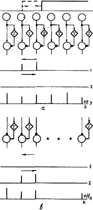
Fig. 2. A contour detection diagram. Top: The “on” diagram; bottom: the “off” one. Open circles integrating neurons. Delaying neurons are marked by diamonds. Arrows indicate excitatory synapses; filled circles, inhibitory synapses. 1, 2, responses of corresponding neurons to leading or trailing edges; 3, on- and off- responses to illumination of the whole retina.
Skaityti daugiauA spherical model of colour vision, colour discriminability and simultaneous contrast.
Demo: Two colours interaction.
Description: